Urinary tract infection or UTI is one of the most common bacterial infections in childhood. It can be divided into three categories: the upper urinary tract referred to as pyelonephritis, the lower urinary tract, referred to as cystitis, and asymptomatic bacteriuria. Note 1

Unfortunately, it may be difficult, if not impossible to distinguish pyelonephritis from cystitis based on clinical symptoms and signs, especially in infants and young children.
At the end of this article, we should be able to answer the following questions:
- How common is UTI in infants and children (Incidence and Prevalence)?
- What is the common presentation of childhood UTI?
- How do you get UTI (Pathophysiology)?
- How do we diagnose UTI?
- When is renal imaging indicated?
- How do we manage childhood UTI?
How Common is UTI in Infants and Children?
During the first year of life, the incidence of UTI is approximately 0.7% in girls and 2.7% in uncircumcised boys. In febrile infants in the first two months of life, the incidence of UTI is roughly 5% in girls and 20% in uncircumcised boys. During the first six months, uncircumcised boys have a 10 to 12-fold increased risk of developing UTI. In the neonatal period, UTI is more common in premature infants than term infants. After one year of age, girls are much more likely than boys to develop UTIs.

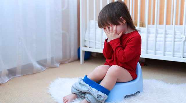
UTI has a bimodal age of onset with one peak in the first year of life, and another peak between two and four years of age which corresponds to the age of toilet training, it has been estimated that approximately 7.8% of girls and 1.7% of boys by the age of seven years will have had a UTI. By the age of 16 years 11.3% of girls and 3.6% of boys will have had a UTI.
A meta-analysis study was conducted to determine the pooled prevalence of urinary tract infection (UTI) in children by age, gender, race, and circumcision status.
* Conclusion: Prevalence rates of UTI varied by age, gender, race, and circumcision status. Uncircumcised male infants less than 3 months of age and females less than 12 months of age had the highest baseline prevalence of UTI. Prevalence estimates can help clinicians make informed decisions regarding diagnostic testing in children presenting with signs and symptoms of urinary tract infection. Note 2
What is the Common Presentation of Childhood UTI?
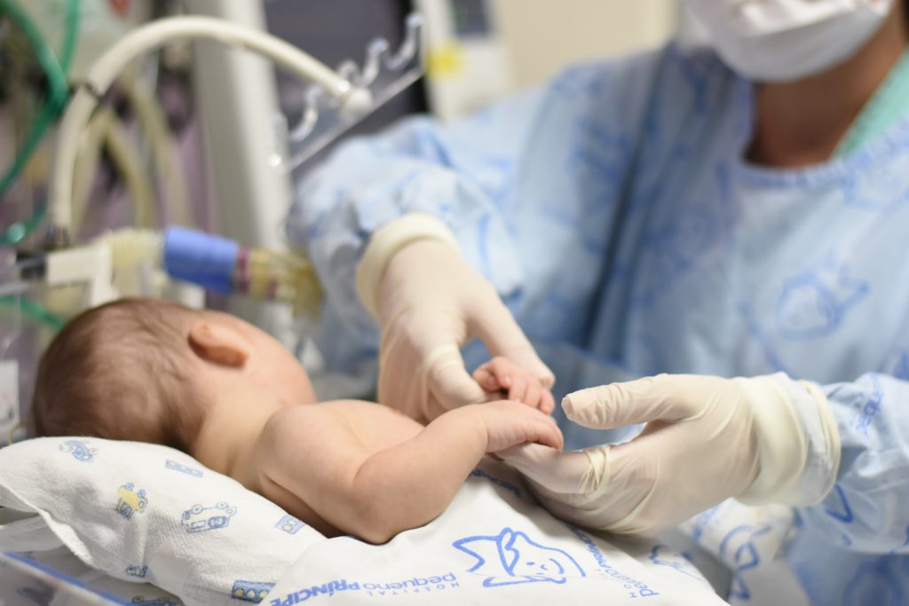
In the neonatal period, the symptoms and signs are non-specific, a neonate might present with signs of sepsis such as:
- Temperature instability
- Peripheral circulatory failure
- Lethargy
- Irritability
- Apnea
- Seizure or metabolic acidosis

Alternatively, a neonate might present with:
- Anorexia
- Poor sucking
- Vomiting
- Suboptimal weight gain or
- Prolonged jaundice
- Foul-smelling urine is an uncommon but more specific symptom of UTI
- Septic shock is unusual unless the patient is compromised or obstruction is present
- In neonates, with UTI there is a high probability of bacteremia suggesting the hematogenous spread of the bacteria.
The symptoms of UTI usually remain non-specific throughout infancy, unexplained fever is the most common during the first two years of life, in fact, it may be the only presenting symptom of UTI in this age group.

In general, the prevalence of UTI is greater in infants with temperatures greater than 39 degrees centigrade than those with temperatures less than 39 degrees centigrade.
Other non-specific manifestations include:
- Irritability
- Poor feeding
- Anorexia
- Vomiting
- Recurrent abdominal pain and
- Failure to thrive
Specific symptoms and signs include:
- Increased or decreased number of wet diapers
- Malodorous urine and discomfort with urination
- A weak or dripping urinary stream suggests a neurogenic bladder or obstruction in the low urinary tract such as posterior urethral valves in boys
- Constant dripping of urine or wetting of diapers may suggest the presence of an ectopic ureater, a predisposing factor to UTI.
After the second year of life, the symptoms and signs of UTI are more specific symptoms and signs of pyelonephritis include:
- Fever
- Chills
- Rigor
- Vomiting
- Malaise
- Flank pain
- Back pain and
- Costo vertebral angle tenderness
Lower tract symptoms and science include:
- Suprapubic pain
- Abdominal pain
- Disuria urinary frequency
- Urgency
- Cloudy urine
- Malodorous urine
- Daytime wetting
- Nocturnal and euresis of recent onset and
- Suprapubic tenderness
Urethritis without cystitis may present as sharia without urinary frequency or urgency.
What are the Common Pathogens Causing UTIs?
The most common causative organisms are from the intestinal flora:
- Escherichia coli accounts for 80 to 90% of UTIs in children Note 1
- Enterobacter aerogenes
- Klebsiella pneumoniae
- Proteus mirabilis = more common in boys than in girls
- Citrobacter
- Pseudomonas aeruginosa
- Enterococcus spp.
- Serratia spp.
- Streptococcus agalactiae is relatively more common in newborn infants.
- Staphylococcus saprophyticus is very common in sexually active female adolescents, accounting for 15% of UTI.
- In children with anomalies of the urinary tract (anatomic, neurologic, or functional) or compromised immune system. staphylococcus aureus, staphylococcus epidermidis, haemophilus influenzae, streptococcus pneumoniae, streptococcus viridians, and streptococcus agalactiae may be responsible.
- Hematogenous spread of infection an uncommon cause of UTI may be caused by staphylococcus aureus, streptococcus agalactiae, proteus mirabilis, pseudomonas aeruginosa, and non-typhoidal salmonella.
- Rare bacterial causes of UTI include mycobacterium tuberculosis, and streptococcus pneumoniae.
Viruses such as adenoviruses, enteroviruses, echoviruses and coxsackieviruses may cause UTI. The associated infection is usually limited to the lower urinary tract. In this regard adenoviruses are known to cause hemorrhagic cystitis.
Fungi such as candida spp, cryptococcus neoformans and aspergillus spp are uncommon causes of UTI, and occur mainly in children with an indwelling urinary catheter, anomalies of the urinary tract, long-term use of broad-spectrum antibiotic, or compromised immune system.
The predisposing factors for the development of UTI in infants and children include:
- Lack of breastfeeding
- Umale
- Hypercalceuria
- CAKUT (congenital anomalies of the kidney and the urinary tract) such as vur, upg, puv Note 3
- Bladder dysfunction
- Constipation
- Sexual intercourse
An important predisposing factor in childhood UTI is CAKUT. Prevalence of structural abnormalities in children with UTI ranges from 10 to 75%. Risk of UTI in children with specific structural abnormalities range from 15 to 50%.
The pathogens reach the urinary tract in three ways:
- The majority 91 to 96% of UTI results from the ascent of bacteria from the periurethral area, migrating in a retrograde fashion via the urethra to reach the bladder and potentially the upper urinary tract. Periurethral colonization with uropathogenic bacteria is considered an important factor.
- Hematogenous spread can also occur and is more common in the first few months of life.
- Bacteria may also be introduced into the urinary tract via instrumentation, such as catheterization.
The development of UTI is an interplay of host behavior characteristics and virulence factors of the pathogen.
The increased susceptibility of girls to UTI might be explained by the relatively shorter length of the female urethra and the regular heavy colonization of the perineum by enteric organisms. Factors that increase colonization of the female perineum include:
- High vaginal pH
- The increased adhesiveness of bacteria to vaginal cells
- Diminished cervical vaginal antibody
The prefucial space is a potential reservoir of bacterial pathogens in boys.
Host behavior that predisposes to UTI includes:
- Voiding postponement
- Infrequent voiding
- Sexual intercourse is an important risk factor in female adolescence
- Poor hygiene
The presence of stone can serve as a nidus for bacteria causing UTI. Urge syndrome and dysfunctional voiding are associated with post-void residual urine which predisposes to UTI. Other adverse host factors include parenchymal renal anomalies, dysfunctional bladder emptying, detrusor muscle instability, constipation, diabetes mellitus, immunodeficiency, obesity, and vitamin D deficiency, infants especially neonates are at a higher risk for UTI presumably due to their incompletely developed immune systems. Recent studies have shown that children and adolescents with psychosis are more prone to UTI.
Local bladder wall defense mechanisms such as through the production of mucus and secretion of antimicrobial peptides by the uroepithelium, restrict attachment of bacteria to uroepithelial cells. In addition, the uroepithelium expresses toll-like receptors with the capacity to recognize pathogen-associated molecule patterns. Engagement of toll-like receptors can lead to uroepithelial cell activation and production of inflammatory mediators such as cytokines which generate a local inflammatory response to facilitate the eradication of the invading bacteria. Regular voiding with the anti-grade flow of urine and complete bladder emptying minimize the opportunity for attachment. The urethral flora includes aerobic bacteria. However, the growth of these micro-organisms is inhibited by low urine pH, soluble IgA, polymorph nuclear cells, lactoferrin, lipocalin, Tamm-Horsefall glycoprotein, and a high organic acid or urea concentration in the urine.
Virulence factors of pathogens increase the likelihood that a specific bacterial strain will colonize and subsequently invade the urinary tract. These factors include:
- Hemolysin
- Hemagglutinin
- Endotoxin
- Cytotoxic necrotizing factor 1
- K capsular antigen
- A rigid cell wall
- Serum resistance ability due to the outer membrane protein TraT
- Aerobactin which supports growth by chelating iron
- Adhesive capacity
The three different types of adhesins identified on uropathogenic E. coli include:
- Type 1 pili (or fimbriae)
- P-fimbri
- X-adheasins
These adhesions facilitate adherence of the bacteria to mucosal receptors in the uroepithelium in spite of the flushing action of urine flow. Once the uroepithelium is invaded, an intracellular biofilm is formed. The biofilm can protect the uropathogenic E. coli from the host immune system.
Conditions that interfere with the uni-directional flow of urine increased susceptibility to UTI. This occurs with vesico ureteric reflux and obstruction. Vesicoureteric reflux the most common urologic anomaly in children allows bacteria to ascend from the bladder to the kidney, and also leads to post-void residual urine. Vesicoureteric reflux can be primary or secondary to the posterior urethral valve, ectopic ureter, or prune belly syndrome. It is an important risk factor for recurrent UTI and renal scarring. Vesicoureteric reflux occurs in 25 to 30% of children who have had a UTI. Primary vesico ureteric reflux often resolves spontaneously while secondary vesico-ureteric reflux only resolves with correction of the underlying cause.
Anatomic obstruction with resultant stasis of urine can occur due to fimosis, metal stenosis, labial fusion, posterior urethral valves, urethral strictures, uroteroseals, ureterovesicle or ureteropelvic junction obstruction, renal stone or extrinsic mass. For example fecal impaction, tumor cyst. These may predispose the child to UTI.
How Should We Collect Urine Specimen in Children?
Collection of urine is a very important component in the diagnosis of UTI. Urine collection in infants and children can be challenging, however there is a need for specimens to be collected properly. There are three methods of collection:
- Suprapubic aspiration
- Transurethral catheterization
- Clean voided samples
Currently there is no consensus regarding the best method for urine collection in children who are not toilet trained. Note 4 The American Academy of Pediatrics recommends that the urine specimen needs to be obtained through suprapubic aspiration or catheterization only. Unfortunately suprapubic aspiration and catheterization are invasive, stressful and may not be feasible in a primary care setting. On the other hand, the National Institute for Health and Clinical Excellence (NICE) and the Italian Society of Pediatric Nephrology (ISPN) recommend the clean-catch method as the method of choice for urine collection, reserving suprapubic aspiration or catheterization for specific situations such as a febrile child in poor general health or appears severely ill. The Canadian Pediatric Society recommends leaving the child with the diaper off and obtaining a clean-catch urine sample when the child voids. If the urinalysis is abnormal, urine collection by catheterization or suprapubic aspiration would be in order.
Suprapubic aspiration is a useful method to obtain a clean urine sample from infants as well as from those children who are incontinent or acutely ill. The procedure is contraindicated in children with coagulopathy or an abdominal wall defect. Since most failures are caused by the absence of urine in the bladder, a suprapubic tap should not be performed in an infant who has recently voided. The success rate is higher when ultrasonographic guidance is used to ensure the bladder is adequately full and allow visualization of structures between the abdominal wall and the bladder. Complications of suprapubic aspiration include transient gross hematuria and the rare inconsequential puncture of other abdominal viscera. Suprapubic aspiration should be reserved for situations in which a clean-catch or catheter specimen is not otherwise readily available or when the time is of essence.
Catheterization of the urinary bladder to obtain a urine specimen for culture is not routinely recommended. Catheterization is associated with discomfort for the child. Emotional stress for both the child and the parents significant trauma with consequent disuria and hematuria and potential introduction of infection into the bladder.
A clean-catchment stream urine specimen obtained after proper cleansing of the external genitalia is satisfactory for most diagnostic purposes in children who can void on demand. During cleansing and voiding, the risk of contamination can be minimized by having young girls sitting backward on the toilet seat and gently spreading the labia in girls and in uncircumcised boys retracting the foreskin. A randomized controlled trial on Quik-Wee method proves that a clean voided sample can be successfully obtained in an infant as well.
How is the clean-catchment method done?
Feed the baby for one hour, then clean the genital orifice with sterile water at room temperature for 10 seconds, place the sterile bag in the proper position, rub the suprapubic area of the child in a circular pattern with gauze soaked in cold saline stored in 2.8 degree centigrade held with disposable plastic forceps then wait until a clean catch urine sample is obtained.
The Quick-Wee method significantly increased the voiding and success rate of clean-catch urine for infants aged 1 to 12 months. This method requires minimal resources and is a simple way to trigger faster voiding for clean-catch urine from infants in the acute care setting. The urine should be collected in a sterile container and the specimen should not be allowed to stand in room air, because the number of bacteria will double every 30 minutes. If the urine cannot be examined microscopically or plated within a short period, the specimen can be refrigerated at four degrees celsius for up to four hours with minimal alteration in the results of the colony count.
What Are the Test Types For Urine Specimen?
The specimen can be sent for dipstick, microscopy and urine culture
Dipstick Tests
Dipstick tests are inexpensive, convenient, readily available and useful for diagnosis of UTI.
The leucocyte esterase dipstick test demonstrates the presence of pyuria by histochemical methods that detect this enzyme and neutrophils. Leucocyte esterase is also present even if leucocytes are lysed. On the other hand, a positive leukocyte esterase dipstick test is not diagnostic as leucocytes may be present in the urine in other conditions such as kawasaki disease, hypercalceuria, gastroenteritis and appendicitis. The leucocyte esterase dipstick test may be falsely negative if leucocytes are present in low concentration. The test has a specificity of approximately 78% and a sensitivity of 83%.
Application of the nitrite test is based on the principle that dietary nitrate in the urine will be reduced to nitrite if large numbers of nitrate-reducing bacteria, for example E coli, klebsiella species, proteus species are present and sufficient time is available to complete the reaction. The nitrite test has a specificity of approximately 98% and a sensitivity of 53%. False negative reactions can result from inadequate dietary nitrate, inadequate time, less than four hours for the conversion of nitrate into nitrite due to frequent emptying of the bladder infection caused by non-nitrate reducing bacteria. For example pseudomonas species, enterococcus species, staphylococcus saprophyticus, antimicrobials that inhibit bacterial metabolism, a large volume of dilute urine or ascorbic acid in the urine. Rapid urine tests have a false negative rate of approximately 10% and therefore cannot replace urine culture in the diagnosis of UTI. Nevertheless rapid urine tests are good screening tests which when used in conjunction with urine culture can increase its diagnostic accuracy.
Microscopy
Microscopy should be performed to detect bacteria urea and pyuria. The presence of crystals or a significant number of urothelial cells, vaginal cells, red blood cells or white blood cells can mask the presence of bacteria in the urine. Gram stain of a urine specimen has a sensitivity of 81% and a specificity of 83%. If a ground stain of uncentrifuged urine specimen shows one or more bacteria per oil immersion field. there is an 80 to 95% correlation with a colony count of greater than 105 cfu/ml. The presence of five white blood cells per high power field in centrifuged urine or 10 white blood cells as detected by hemocytometer in uncentrifuged urine respectively is the gold standard for pyuria.
Pyuria is not diagnostic of UTI. Hyuia has a specificity of approximately 81% and sensitivity of 73%. Sterile pyuria can be associated with infection due to anaerobic bacteria, tuberculosis, viral pathogens, chemical or allergic inflammation, cervical or vaginal secretion, Kawasaki disease, crystal Luria, appendicitis, regional enteritis, glomerulonephritis, and interstitial nephritis. Conversely the absence of pyuria on a single specimen does not rule out UTI. Serial urinalysis in patients with UTI eventually shows pyuria.
Urine Culture
Urine culture remains the gold standard for diagnosing UTI. Bacteria are usually evident in properly plated urine specimens by about 24 hours and sensitivity results are usually available within 48 hours. When a routine culture is negative but the child is symptomatic or the ground stain demonstrates bacteria, an anaerobic culture should be obtained. If the urine culture yields unusual bacteria or multiple bacteria in an uncontaminated urine specimen, immunodeficiency and malformation of the kidney and urinary tract should be excluded. Note 5
Criteria for significant bacteria depends on method of collection as shown in this table:
| Method of Collection | Colony count/ml
(Pure culture) |
Probability of infection (%) |
| Suprapubic Aspiration | Gm-neg bacilli:any
Number Gram-pos cocci: >10³ |
>99% |
| Transurethral Catheterisation | >10⁵
10⁴ – 10⁵ 10³ – 10⁴ <10³ |
95%
Infection likely Suspicious, repeat Infection unlikely |
| Clean void
Boy Girl |
>10⁴
3 specimens >10⁵ 2 specimens> 10⁵ 1 specimen >10⁵ 5×10⁴ – 10⁵ 10⁴ – 5×10⁴ <10⁴ |
Infection Likely
95% 90% 80% Suspicious, repeat Symptomatic: suspicious, repeat Asymptomatic: infection unlikely |
American, Canadian and European guidelines have different criteria for diagnosis based on dipstick, microscopy and urine cultures.
| Dipstick | Microscopy | Culture | |
| American Academy of Pediatrics (AAP)
Clinical practice guidelines ( 2-4 mo) |
Positive dipstick test (leukocyte esterase and/or nitrite test) | Positive for pyuria or bacteriuria | > 50,000cfu/ml of a uropathogen in a catheterized or supra |
| Canadia Paediatric Society (CPS) guidelines | Positive dipstick test (leukocyte esterase and/or nitrite test) | Positive urine culture of a single uropathogen (> 100,000cfu/ml in a midstream urine specimen, > 50,000cfu/ml in a catheterized specimen, and any organism in a suprapubic aspiration specimen) are required for the diagnosis of UTI |
Do All Infants and Children Need Imaging?
In the workup of children with UTI physicians must judiciously utilize imaging studies to minimize exposure of children to radiation. Imaging studies are indicated only for children at risk of developing renal damage. The aim of imaging in children with UTI is to identify those with underlying abnormalities or factors that put them at increased risk of recurrent UTI or renal damage. A child is considered at risk of subsequent complications if the following features are present:
- High fever
- Toxicity
- Poor urinary strain
- Palpable kidneys
- Unusual organism isolated in urine culture (other than E. coli)
- Failure to respond to antibiotic treatment within 48 hours
- Recurrent UTI. Recurrent UTI is defined as two or more episodes of UTI with acute pyelonephritis upper UTI, or one episode of UTI with acute pyelonephritis upper UTI plus one episode of uti with cystitis lower uti, or three or more episodes of UTI with cystitis lower UTI.
Renal and bladder ultrasonography is the method of choice to image the urinary tract. Ultrasonography is non-invasive, safe, easy to perform and radiation free. With renal and bladder ultrasound, hydration is essential to sufficiently fill the bladder filling. In toilet trained children, a post-void evaluation is essential.
A renal and bladder ultrasound can define the kidney size, shape and position, echotexture of the renal parenchyma, the presence of duplication and dilatation of the ureters, obstructive uropathy, and structural abnormality of the bladder. The degree of echogenicity and the sharpness of the corticometallary differentiation provide clues to the presence of generalized renal disease or local damage.
Renal ultrasonography can also identify renal or perirenal abscess or pionephrosis.
Renal and bladder ultrasonography should be considered in children two years of age with a febrile UTI, children of any age with recurrent UTI, and children with palpable abdominal mass, abnormal voiding, hypertension, hematuria, no response to standard antimicrobial treatment, and family history of renal or urological disease. Note 6
Ultrasonography is not a sensitive study to localize the infection, or to detect a duplicated collecting system, or vesico ureteric reflux. It is, however, useful in predicting the risk of renal scarring. For acutely sick children and children who do not respond to standard antimicrobial treatment, renal and bladder ultrasonography should be performed as soon as possible to rule out urinary tract obstruction or renal abscess, otherwise the procedure should be performed one to two weeks later to reduce the risk of false positive results secondary to renal inflammation.
Renal imaging with 99m Tc-Dimercaptainsuccinic Acid (DMSA) can be used to detect acute pyelonephritis and renal scarring. Decreased renal uptake of the isotope suggests acute pyelonephritis or renal scarring. In addition, a DMSA renal skin can detect the majority 70% of children with moderate to severe vesicoureteric reflux. The routine uses of this imaging modality is not generally recommended due to the radiation hazard and the cost involved.
The NICE guidelines recommend DMSA renal scan four to six months after a typical UTI in children under three years of age, and recurrent UTI in children of any age. It has been suggested that a normal DMSA renal scan may obviate the need for voiding cysto urethrogram in children with recurrent UTI.
If a DMSA renal scan has to be performed, it should be performed in the acute phase to detect acute pyelonephritis, or four to six months later to assess for renal scarring.
Pyelonephritis and urinary tract malformations form a major cause of chronic renal failure. Renal scar develops in up to 5% of girls and 13% of boys after their first symptomatic episode of pyelonephritis. The first two years of life are considered an especially vulnerable time for scarring, with diminishing risk until about eight years of age beyond which the risk is much reduced.
Other factors that predisposed to renal scarring include:
- Pyelonephritis in infancy
- Increased number of pyelonephritic attacks
- Delay of antibiotic treatment
- Bacterial virulence
- Individual susceptibility
Predictors of renal scarring after a first UTI include:
- Temperature over 39 degrees celsius
- Vesico ureteric reflux (especially high-grade)
- Bbnormal renal/bladder ultrasonography
- Increased absolute neutrophil count
- Increased serum procalcitonin
- C-reactive protein greater than 40 mg/l
- TGF-β1 gene polymorphisms
Approximately 10% of children with a renal scar will develop hypertension in adolescence or early adulthood. Females with a renal scar are at increased risk for toxemia in pregnancy. Renal insufficiency and end-stage renal disease is a possible consequence of renal scarring from pyelonephritis. Complications such as renal abscess, pionephrosis, emphysema II pyelonephritis, and xanthogranulometas pyelonephritis are rare in the post-antibiotic era. It has been shown that 25 to 30% of children with UTI have vesico-ureteric reflux.
A voiding cystourethrogram is the preferred screening test for vesicoureteric reflux. This study accurately grades vesicoureteric reflux, identifies posterior urethral valves, ureteroceles, obstructive uropathies, and other abnormalities of the urethra, ureter, and bladder (e.g. bladder diverticuli or trabeculations), and provides clues to the presence of urge syndrome and dysfunctional voiding.
The procedure is invasive and costly and exposes the child to radiation. A voiding cystourethrogram does not need to be performed after the first febrile UTI unless renal and bladder ultrasonography reveals scarring, hydronephrosis or other findings suggestive of high-grade vesico-ureteric reflux, or obstructive uropathy. The voiding cystourethrogram is also not required after the first febrile UTI and other atypical or complex clinical circumstances, such as poor growth hypertension and a combination of high fever (temperature over 39 degrees celsius) and a pathogen other than E. coli. This is because only a small number of children with vesico-ureteric reflux ultimately require medical or surgical treatment for the vesico-ureteric reflux. Otherwise the procedure should be performed if the child has two or more febrile UTI. Traditionally a voiding cystourethrogram is usually scheduled several weeks after the febrile UTI. It is now recognized that the procedure may be performed as long as the patient is asymptomatic. The risk of post-procedural UTI after a voiding cystourethrogram is very low.
What Are the Possible Complications of UTI?
Renal insufficiency is a well-known complication, either from pyelonephritis per se, a pre-existing congenital renal anomaly which predisposes the child to UTI, or from the use of nephrotoxic antibiotics.
UTI distresses the child, concerns the parents and is a common cause of discomfort. In addition to missed school and work, the condition may adversely affect the quality of life of the child or parents, especially if the UTI is recurrent, or causes permanent renal damage. UTI in infancy is a risk factor for recurrent abdominal pain in childhood.
What Should You Do?
Children should be instructed to void about every 1.5 to t2 hours and never to hold the urine to the last minute. With voiding, children should be encouraged to use optimal posture, and take time to completely empty themselves. Meticulous genital hygiene and adequate fluid intake should be encouraged. Underlying conditions such as constipation, dysfunctional voiding should be treated.
Prompt antibiotic therapy is indicated for symptomatic UTI based on clinical findings and positive urinalysis while waiting for the culture results to eradicate the infection and improve clinical outcome.
The empiric antibiotic chosen should provide adequate coverage for Gram-negative rods notably E. coli and Gram-positive Cocci.
The ideal antibiotic should be easy to administer, achieve a high concentration in the urine, have minimal or no effect on the fecal or vaginal flora, have a low incidence of bacterial resistance, have minimal or no toxicity, and have a low cost.
